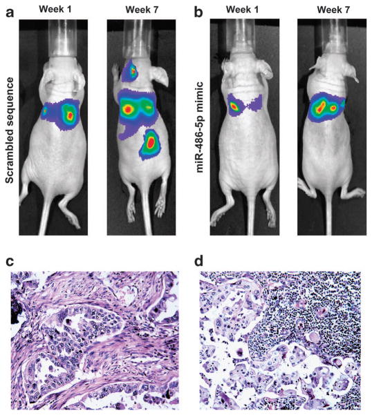
Figure 3.
Forced expression of miR-486-5p restrains NSCLC metastasis in vivo. (a) Fluorescent imaging analysis showed that malignant lesions in the lungs of the mice injected with H460-luc2 cells transfected with scrambled sequence after 1 week, in the neck and abdomen after 7 weeks. Ten mice were used in the group. All 10 mice were monitored by using fluorescent imaging weekly for tumor formation. The figure only shows images that were taken at week 1 and week 7 from the same mouse. (b) Small positive imaging signal was obtained in the lungs of the mice injected with H460-luc2 cells transfected with miR-486-5p mimic after 1 week, and no positive signal in the neck and lower abdomen after 7 weeks. Ten mice were used in the group and were monitored using fluorescent imaging weekly. The figure only shows images that were taken from the same mouse at week 1 and week 7. More description about the data obtained from all 10 mice from each group was shown in Supplementary Figures 4–6. (c) The mice were killed and autopsied in week 8 after cancer cell injection. Serial sections were made from the lungs and metastatic tumor tissues and stained with hematoxylin and eosin (H&E). H&E staining confirms that the cancer cells transfected with scrambled sequence produced tumor in the lungs. (d) H&E staining shows that H460 cells with scrambled sequence created a metastatic tumor in lymph node.

