Fig. 2.

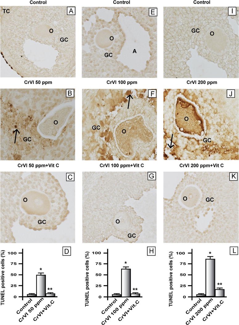
Effect of lactational exposure to CrIII on apoptosis of granulosa cells in F1 offspring. Lactating mother rats received 50, 100, or 200 ppm CrVI in drinking water, with or without vitamin C supplementation through gavage. Suckling F1 female offspring received Cr through their mother’s milk from PND 1 to 21. On PND 25 the F1 offspring were euthanized and ovaries collected, fixed in 4% paraformaldehyde, embedded, and sectioned. Apoptotic, TUNEL-positive cells were stained brown. A representative image for each treatment group is shown. (A–C) Representative images of ovaries: (A) control PND 25 rats that received no treatment, (B) CrVI 50 ppm, and (C) CrVI 50 ppm+vitamin C supplementation. (D) Histogram showing percentage of TUNEL-positive cells from (A), (B), and (C). (E–G) Representative images of ovaries: (E) control PND 25 rats that received no treatment, (F) CrVI 100 ppm, and (G) CrVI 100 ppm+vitamin C supplementation. (H) Histogram showing percentage of TUNEL-positive cells from (E), (F), and (G). (I–K) Representative images of ovaries: (I) control PND 25 rats that received no treatment, (J) CrVI 200 ppm, and (K) CrVI 200 ppm+vitamin C supplementation. (L) Histogram showing percentage of TUNEL-positive cells from (I), (J), and (K). O, oocyte; GC, granulosa cell; TC, theca cell; A, antral cavity. Arrows indicate apoptotic cells. *Control vs CrVI, **CrVI vs CrVI+vitamin C, P<0.05.
