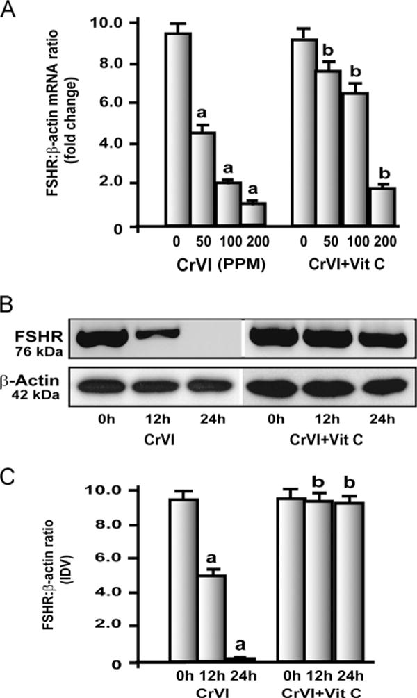
Fig. 3.

Effects of CrIII on FSH-receptor (FSHR) mRNA expression in the ovary. Experimental details are described under Materials and methods. (A) FSHR mRNA was quantified by real-time PCR in the ovary of rats that were exposed to CrIII through mother’s milk between PND 1 and 21. (B, C) Effects of CrVI treatment on FSHR protein expression in primary cultures of granulosa cells (GC). In vitro experiments were carried out in triplicates in the six groups as follows: control, CrVI 12 h, CrVI 24 h, vitamin C, CrVI 12 h+vitamin C, and CrVI 24h+vitamin C. Cells were treated with 10 μM potassium dichromate with or without pretreatment with 1 mM vitamin C. Expression of FSHR protein was quantified by Western blot analysis. (B) Representative immunoblots of FSHR and β-actin proteins and (C) histogram showing FSHR:β-actin ratio. CrVI treatment decreased FSHR in GCs in a time-dependent manner, and vitamin C inhibited the effects of CrVI treatment. Each value is the mean±SEM of three different plates per treatment, P<0.05; aCrVI treatment, 0 h vs 12 or 24 h; bCrVI+vitamin C (pretreatment), 0 h vs 12 or 24 h.
