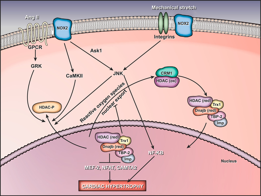
Fig. 2.
Redox signaling pathways regulate cardiac hypertrophy. Nox2 NADPH oxidase stimulation in response to G-protein-coupled receptor agonists or mechanical stretch activates the Ask1–NF-κB pathway, inducing cardiac hypertrophy. HDAC4 suppresses the activity of prohypertrophic transcription factors. Phosphorylation or oxidation of HDAC4 during oxidative stress conditions results in its export to the cytosol, leading to hypertrophy [511]. Abbreviations used: AngII, angiotensin II; Ask1, apoptosis signaling kinase 1; CaMKII, calcium/calmodulin-dependent kinase II; CAMTA2, calmodulin-binding transcription activator 2; CRM1, chromosomal region maintenance-1; Dnajb5, DnaJ homolog subfamily B member 5; GPCR, G-protein-coupled receptor; GRK, G-protein-coupled receptor kinase; HDAC, histone deacetylase; HDAC-P, phosphorylated HDAC; Imp, importin α; JNK, c-Jun N-terminal kinase; MEF-2, myocyte enhancer factor-2; NFAT, nuclear factor of activated T cells; NF-κB, nuclear factor-κB; ox, oxidized; red, reduced; TBP-2, thioredoxin binding protein-2; Trx1, thioredoxin 1.

