Fig. 2.
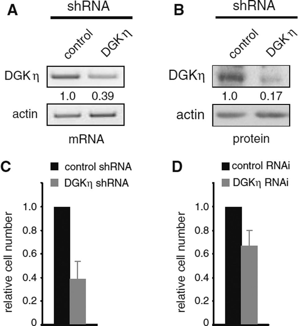
DGKη depletion reduces cell proliferation. H1650 cells were used to generate control or DGKη shRNA polyclonal cell lines. The cells were grown to confluence, harvested, and then DGKη and actin were detected using semi-quantitative RT-PCR (a) or Western blotting (b). The relative levels of DGKη normalized to actin are shown below the DGKη images. c Equal numbers of control or DGKη shRNA H1650 cells were grown for 5 days in medium with 1 % serum and then counted. The difference in cell numbers between control and DGKη shRNA cells was statistically significant (n = 3; p < 0.02). d Control or siRNA oligonucleotides were used for transient depletion of DGKη in H1650 cells grown in 1 % serum. Cell numbers were determined 3–4 days later. The difference in cell number between control and DGKη RNAi cells was statistically significant (n = 3; p < 0.05)

