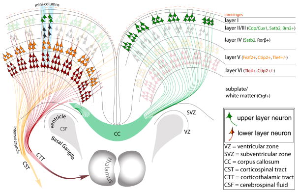
Figure 1. A simplified schematic of the postnatal organization and projections of neocortical projection neurons.
The neocortex is highly organized in both horizontal and vertical dimensions. Horizontally, six layers are defined by highly organized subpopulations of glutamatergic projection neurons, which represent approximately 85% of all neocortical neurons. These subpopulations of projection neurons are characterized by specific molecular identities, dendritic morphologies and terminal targets corresponding to each layer. Projection neurons that are born during later stages of prenatal neurogenesis will be predominantly placed in upper layers II–IV (green neurons). These neurons express specific transcription factors like CDP/Cux1, and project solely intracortically forming the corpus callosum that connects the two hemispheres. However, there is also a smaller portion of intracortically projecting neurons placed in lower layers too (not shown). In contrast, earlier born projection neurons will be placed in lower layers V–VI (orange and red neurons). These subopulations will express transcription factors like FEZF2, and will project subcortically to form long range tracts across the central nervous system like the corticothalamic tract (CTT) originating mainly from layer 6, and somewhat from layer 5, or corticospinal tract (CST) originating solely from layer 5. Within the subventricular zone (SVZ) of the corticostriatal junction, adult progenitors are found giving rise to olfactory cortex neurons.

