Fig. 3.

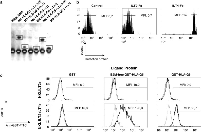
Recognition of the HLA-G α1–α3 structure by LILRB1 and LILRB2. a Immunoprecipitation of HLA-G1 and HLA-G5 (α1–α2–α3 domains), and HLA-G2 and HLA-G6 (α1–α3 domains) with LILRB2-Fc. b Differential direct binding of HLA-G6-GST recombinant protein to LILRB1-Fc and LILRB2-Fc-coated beads. c Differential binding of B2M-free HLA-G5-GST and HLA-G6-GST recombinant proteins to NKL-LILRB1+ and NKL-LILRB1+LILRB2+ cells by flow cytometry analysis, using GST recombinant protein as control and anti-GST antibody for detection
