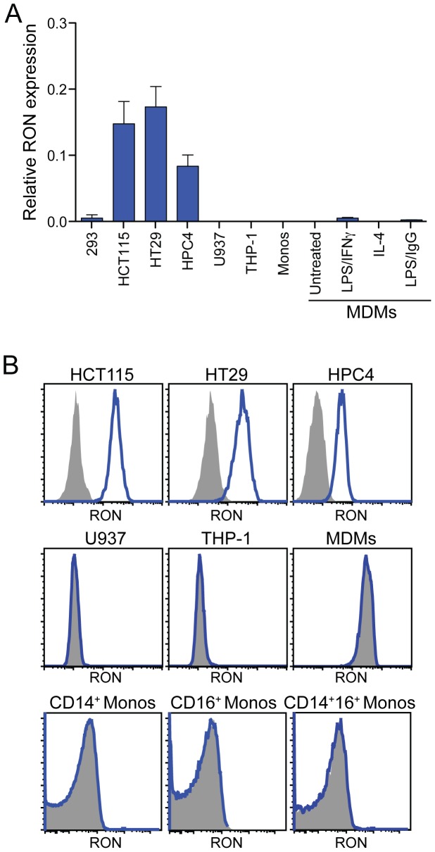
Figure 2. RON is preferentially expressed by human epithelial cells.
(A) Quantitative RT-PCR survey of RON expression in human cell lines and primary cells. Columns 1: HEK293 control, columns 2–4: epithelial cell lines, columns 5–6: monocytic cell lines, column 7: human CD14+ monocytes (monos), columns 8–11: monocyte-derived macrophages (MDMs) left untreated or treated with the indicated stimuli. Data in columns 1-7 are the mean +/− SD of three independent samples and columns 8–11 are the mean of two donors. (B) Analysis of single-cell suspensions for RON expression by flow cytometry. Cells were stained with a monoclonal antibody specific for human RON (blue histogram) or an isotype control antibody (shaded histogram). MDMs were gated as CD14+CD33+.

