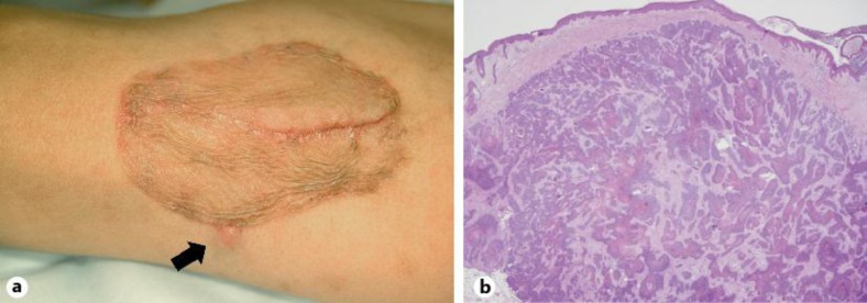
Fig. 3.
Clinical and histopathological features illustrating the local recurrent lesion. a Clinical photograph of the subcutaneous induration on the periphery of the skin graft (arrow: Case 4). b Histopathology of the recurrent lesion demonstrated intradermal proliferation of EPC cells (Case 4; HE; ×20).

