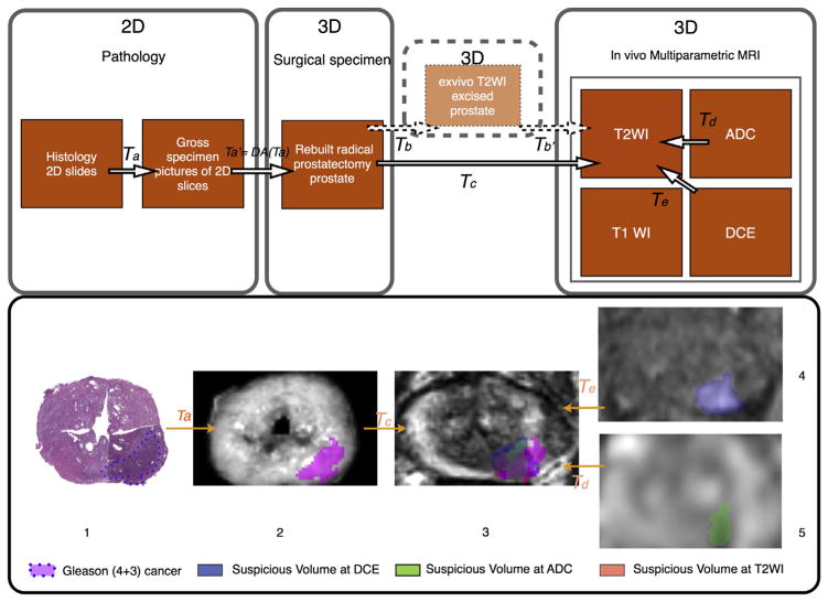
Figure 2.
Co-registration workflow applied to cancer focus. 1st row. Workflow for pathology–mpMRI co-registration in a surgical 3D space. 2nd row. 3D deformable co-registration of virtual whole-mount histology (1), fresh specimen (2), T2WI (3), perfusion (4), and diffusion (5) sequences (ADC) applied to prostate cancer Gleason score 7 (4 + 3). The T2WI is the common space. Data extracted from the 3D volume (2,3,4,5). mpMRI, multiparametric magnetic resonance imaging; T2WI, T2-weighted imaging; ADC, apparent diffusion coefficient; T1WI, T1-weighted imaging; DCE, dynamic contrast-enhanced weighted images; 2D, two-dimensional 3D, three-dimensional; T(x), transformation x.

