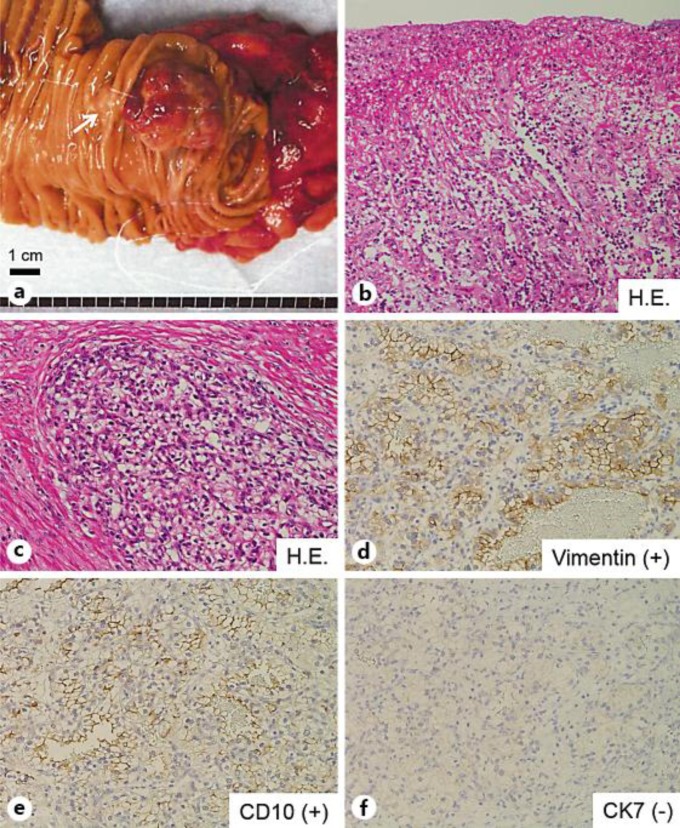
Fig. 2.
a Macroscopic findings of the resected specimen reveal a polypoid mass in the descending portion of the duodenum that appears ulcerative and friable. The papilla of Vater is indicated by an arrow. b Histologic findings show that the surface of the tumor was coated by granulation tissue consisting of inflammatory cells, fibrosis and edematous stroma. c Histologic image shows dysplastic clear cells containing glycogen and arranged in an alveolar pattern. d–f Immunohistochemical staining demonstrates that the clear cells are positive for vimentin (d) and CD10 (e) and negative for CK7 (f), confirming the diagnosis of RCC with clear cell histology.

