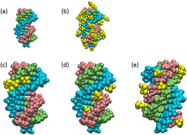
Figure 4. Estimation of Binding of Choline ions to the triplex by MD simulations.
A snapshot of Ts1 after 20 ns MD simulation in (a) the absence and (b) presence of choline ions. First (5′-TTTTTTTCTTCT-3′), second (5′-AGAAGAAAAAAA-3′) and third (5′-TCTTCTTTTTTT-3′) strands in Ts1 are indicated by light blue, light green and pink, respectively. Ts1 and choline ions (yellow) are depicted as van der Waals models. The choline ions bound to minor and ma-major grooves in Ts1 are highlighted in (c) and (d), repectively. The choline ions surrounding the third strand in Ts1 are highlighted in (e).

