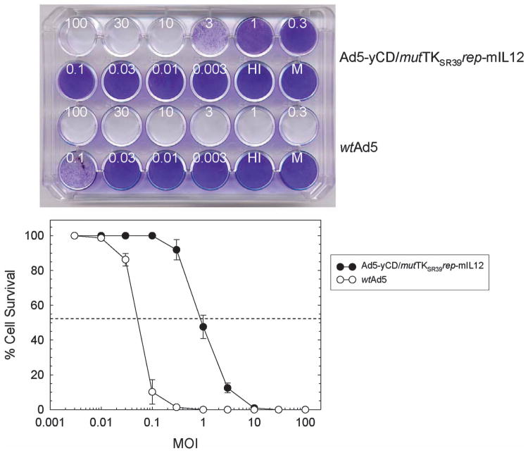
Figure 2.
CPE assay. DU145 cells were infected with varying amounts (in PFU per cell) of Ad5-yCD/mutTKSR39rep-mIL12 or wild-type Ad5 (wtAd5). Cells were fixed and stained with crystal violet 7 days later (top), and cell survival was quantified (bottom). The results represent the mean of triplicate determinations ± s.d. HI, heat-inactivated adenovirus; M, mock infection.

