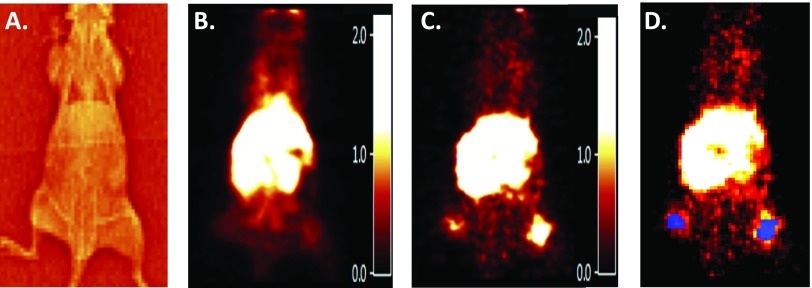
Figure 2.
[11C]-erlotinib imaging of a mouse bearing bilateral HCC827 and PC9 bilateral flank xenografts. (A) Transmission scan of the mouse used to gain anatomic information for ROI placement. (B) [11C]-erlotinib PET early time image summed from 0 to 10 minutes. PET images are smoothed with a 1.0-mm3 kernel for illustrative purposes. (C) [11C]-erlotinib PET late time image summed from 95 to 120 minutes. (D) ROIs (blue) corresponding to flank xenograft location used for data acquisition and TAC analysis.

