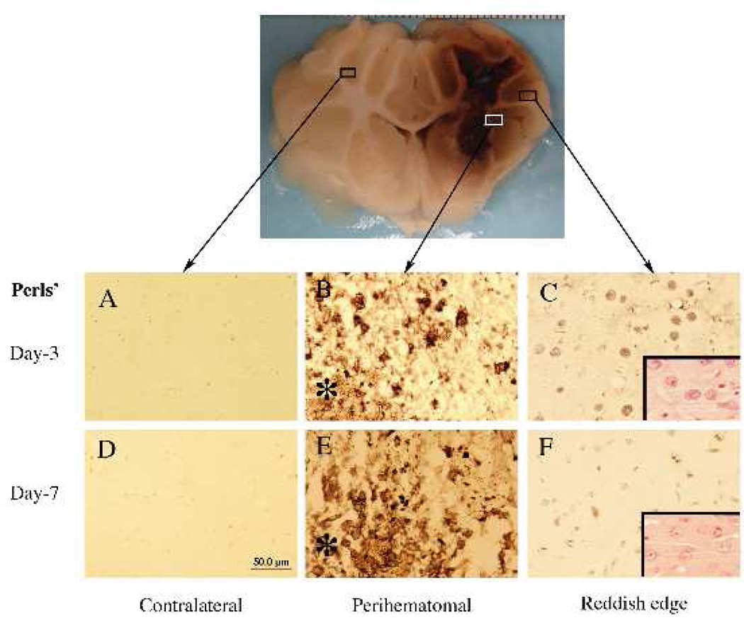
Figure 5.
Iron histochemistry (Perls’ staining) in the brain 3 days after intracerebral haemorrhage in pigs. Asterisk indicates the haematoma. Insets in C and F: hematoxylin and eosin staining.
Scale bar (A–F)=50 µm. Figure reprinted with permission from Gu et al., Stroke, 2009;40:2241–2243.112

