
Figure 1.
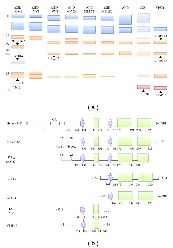
(a) Schematic representation of the spectrum of PrPres fragments observed in human prion diseases and their electrophoretic profile. The unglycosylated forms of all PrPres fragments with the glycosylation sites in their sequence are indicated in orange, while the fragments lacking these sites are shown in red. Among the glycosylated peptides, only the mono- and the diglycosylated forms of PrPres 27–30 (18–21 kDa range) fragments are shown (in blue). The DCF16 fragment, which is generated only in partially denaturing conditions is labeled with a dotted line and a gray color. For GSS, the fragments that have been described only associated with specific PRNP mutations (e.g., P102L or A117V) are shown with dotted lines and in transparency. Molecular weights are indicated on the left in kDa. (b) Diagrams of the secondary structural elements of human PrPC and of the PrPres fragments observed in human prion diseases. Arrows are representative of β-strands and rectangles of α-helices and OR indicates the octapeptide repeats region. The secondary structure numbering has been derived from pdb (Protein Data Bank) id 2LSB (human PrP).