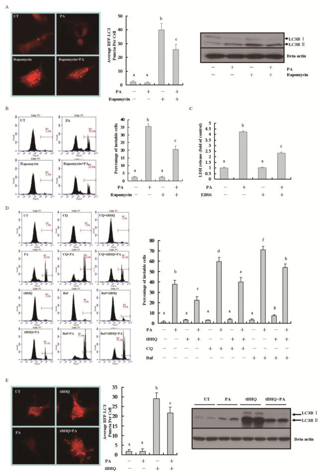
Figure 3.
Autophagy induction contributes to the protective effect of tBHQ on palmitate-induced cell death. (A) AML-12 hepatocytes were treated with 0.5 mM palmitic acid (PA) for 12 h with or without rapamycin pre-treatment for 1 h. For LC3 puncta analysis, 2 × 10⁵/ml cells were seeded into the chamber (BD Falcon, CA) and transfected with mRFP-GFP-LC3 plasmid before palmitic acid (PA) treatment. LC3 puncta formation was examined by fluorescence microscopy. For Western blotting detection, total cell lysates were subjected to immunoblotting assay for LC3B. All values are denoted as means ± SD from three or more independent batches of cells. Bars with different characters differ significantly, p < 0.05. (B) Cell death was detected by propidium iodide staining. All values are denoted as means ± SD from three or more independent batches of cells. Bars with different characters differ significantly, p < 0.05. (C) AML-12 hepatocytes were treated with 0.5 mM palmitic acid (PA) for 12 h in either regular or EBSS medium. Cell death was analyzed by LDH release. All values are denoted as means ± SD from three or more independent batches of cells. Bars with different characters differ significantly, p < 0.05. (D) AML-12 cells were treated with 0.5 mM pamitate acid (PA) for 12 h with or without 1-h pre-incubation with tBHQ (50 μM). Autophagy inhibitors, CQ (20 μM) or Baf (100 nM), were added 1 h before tBHQ addition. Cell death was detected by propidium iodide staining. All values are denoted as means ± SD from three or more independent batches of cells. Bars with different characters differ significantly, p < 0.05. (E) AML-12 cells were treated with 0.5 mM PA for 12 h. tBHQ (50 μM) was added 1 h before PA treatment. For puncta analysis, cells were transfected with mRFP-GFP-LC3 plasmid before 0.5 mM palmitic acid (PA) treatment, and examined by fluorescence microscopy. For Western blotting detection, total cell lysates were subjected to immunoblotting assay for LC3B. All values are denoted as means ± SD from three or more independent batches of cells. Bars with different characters differ significantly, p < 0.05.

