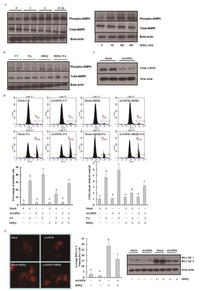
Figure 7.
AMPK activation contributes to tBHQ-triggered autophagy induction. (A) AML-12 cells were treated with tBHQ (50 μM) for indicated time period or for 12 hours at indicated dose. Whole cell lysates were subjected to Western blot for AMPK. (B) AML-12 cells were treated with 0.5 mM palmitate (PA) for 12 h with or without 1 h tBHQ (50 μM) pretreatment. Whole cell lysates were subjected to Western blot for AMPK. (C) AML-12 cells were transfected with either scrambled or AMPK siRNA. Whole cell lysates were subjected to Western blot for AMPK. (D) AML-12 cells were transfected with either scrambled or AMPK siRNA before tBHQ (50 μM) and/or palmitic acid (PA) exposure. Cell death was analyzed by detecting LDH release and propidium iodide staining 12 h later. All values are denoted as means ± SD from three or more independent batches of cells. Bars with different characters differ significantly, p < 0.05. (E) AML-12 cells were co-transfected with mRFP-GFP-LC3 plasmid and AMPK siRNA or scrambled siRNA prior to tBHQ (50 μM) exposure. Puncta formation and LC3B-II conversion were determined by fluorescence microscopy and Western blot, respectively. All values are denoted as means ± SD from three or more independent batches of cells. Bars with different characters differ significantly, p < 0.05.

