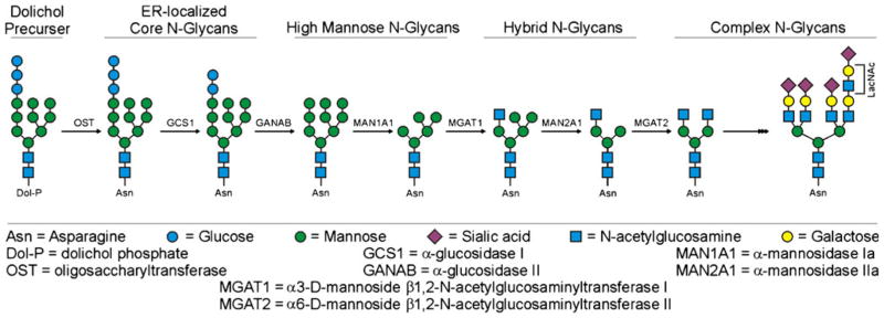
Fig. 1.

Simplified schematic of the N-glycosylation pathway in mammalian cells. Shown are the different classes of N-glycans found in mammals and some, but not all, of the key enzymes that create these structures for the readers easy reference. While this figure depicts events in a particular order, it must also be noted that the pathway is more flexible than shown and some of the enzymes can act in different order. For example, the MGAT1/MAN2A1 reactions can occur in reverse order from what is shown
