Figure 5.
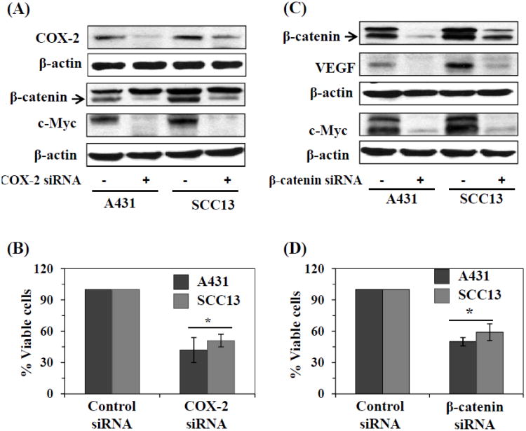
Regulation of β-catenin in human skin cancer cells. (A) siRNA knockdown of COX-2 in A431 and SCC13 cells resulted in decrease in levels of β-catenin and its downstream target c-Myc, as determined by western blot analysis. (B) siRNA knockdown of COX-2 in A431 or SCC13 cells resulted in significant reduction in cell viability. (C) siRNA knockdown of β-catenin in A431 and SCC13 cells resulted in lowering the levels of VEGF and c-Myc, as analyzed by western blot analysis. (D) siRNA knockdown of β-catenin in A431 and SCC13 cells resulted in significant decrease in cell viability. Briefly, for the analysis of cell viability, 5×10⁴ cells were plated in six well culture plates. Twenty-four h later, cells were harvested, counted using a microscope and the cell numbers were compared between control siRNA and β-catenin or COX-2 siRNA knockdown groups of cells. Cells treated with scrambled siRNA were used as a control group. Significant inhibition vs control, *P<0.01.

