FIGURE 7.
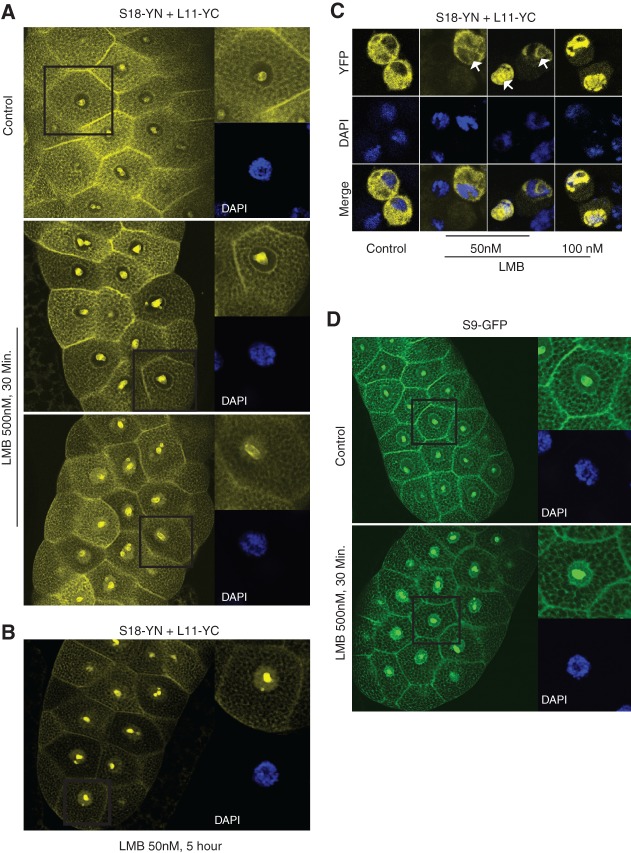
Leptomycin B treatment increases nuclear 80S signal. (A) Large panels are confocal images showing YFP fluorescence in salivary glands incubated for 30 min either without (top panel) or with (bottom panels) 50 nM LMB. Magnified insets show single cells and DAPI signal (blue). (B) Images of salivary glands incubated for 5 h in LMB. (C) Confocal images of S2 cells transfected with the indicated constructs and incubated as indicated with LMB for either 4 h (second panel from left) or 5 h. Arrows indicate nucleoli. (D) Confocal imaging of glands expressing S9–GFP incubated in vitro for 30 min with or without LMB as indicated.

