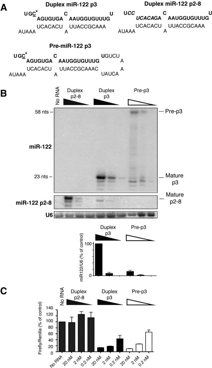
FIGURE 1.
Processing and activities of synthetic miR-122 forms after transfection into HeLa cells. (A) Sequence and predicted structure of duplex p3 and pre-p3. The sequence of mature miR-122 is highlighted in bold. The mutated C-nucleotide at position 3 in miR-122 is underlined and marked with an asterisk. (B) Northern blot to visualize transfected miR-122 RNA species. Duplex p2-8 is a mutated miR-122 duplex with mutations in the seed sequence at positions 2–8. Duplex p3 is a phosphorylated miR-122 duplex with a single mutation at position 3. Pre-p3 is a phosphorylated pre-miR-122 with the same single mutation. Visualization of miR-122 p2-8 was accomplished using a miR-122 p2-8–specific hybridization probe (middle panel). Accumulation of 20- to 23-nt miR-122 bands was normalized to U6 snRNA, and abundance was measured using ImageQuant. (C) Luciferase-based microRNA assays. miR-122 species were transfected into HeLa cells concurrently with a DNA plasmid that expressed firefly luciferase mRNA, containing a miR-122 binding site, and a plasmid that expressed Renilla luciferase. Lysates were prepared and luciferase activities measured. Data from mock-transfected cells (no RNA) were set to 100. Data are representative of at least three independent replicates; error bars, SEM.

