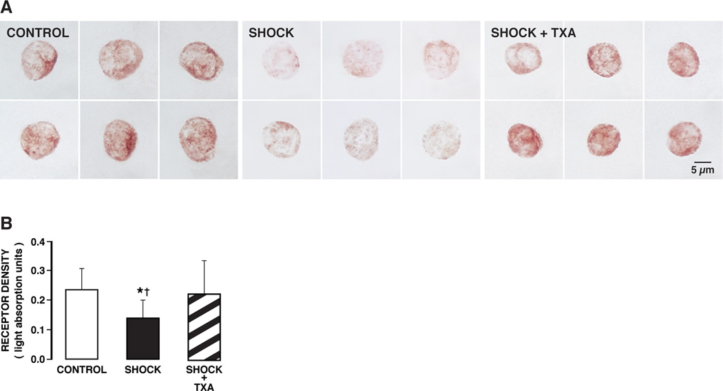
Figure 2.

(A) Brightfield micrographs of formalin-fixed leukocytes immunolabeled with antibody against the extracellular α-domain of the insulin receptor with Vector NovaRED substrate in control animals (CONTROL) and 2 hours after hemorrhagic shock without (SHOCK) and with (SHOCK + TXA) pancreatic digestive enzyme inhibition. The density of the insulin receptor in control leukocytes shows uniform staining of leukocytes as indicated by Vector NovaRED substrate (left panel). The label density was reduced after hemorrhagic shock (middle panel). Hemorrhagic shock animals treated with pancreatic digestive enzyme inhibitor (SHOCK + TXA) (right panel) had insulin receptor densities in leukocytes comparable to control animals (left panel). (B) Light absorption values of the insulin receptor alpha in control animals and after hemorrhagic shock without (Go-Lytely) and with (SHOCK + TXA) pancreatic enzyme inhibition. The average light absorption values of the insulin receptor over randomly selected leukocytes were significantly higher in control and treated animals than without treatment. Pancreatic digestive enzyme blockade with TXA improves extracellular insulin receptor density after hemorrhagic shock. (n = 90 cells/group with 3 rats per group) *p < 0.05 SHOCK vs SHOCK+ TXA, †p < 0.05 SHOCK vs Control.
