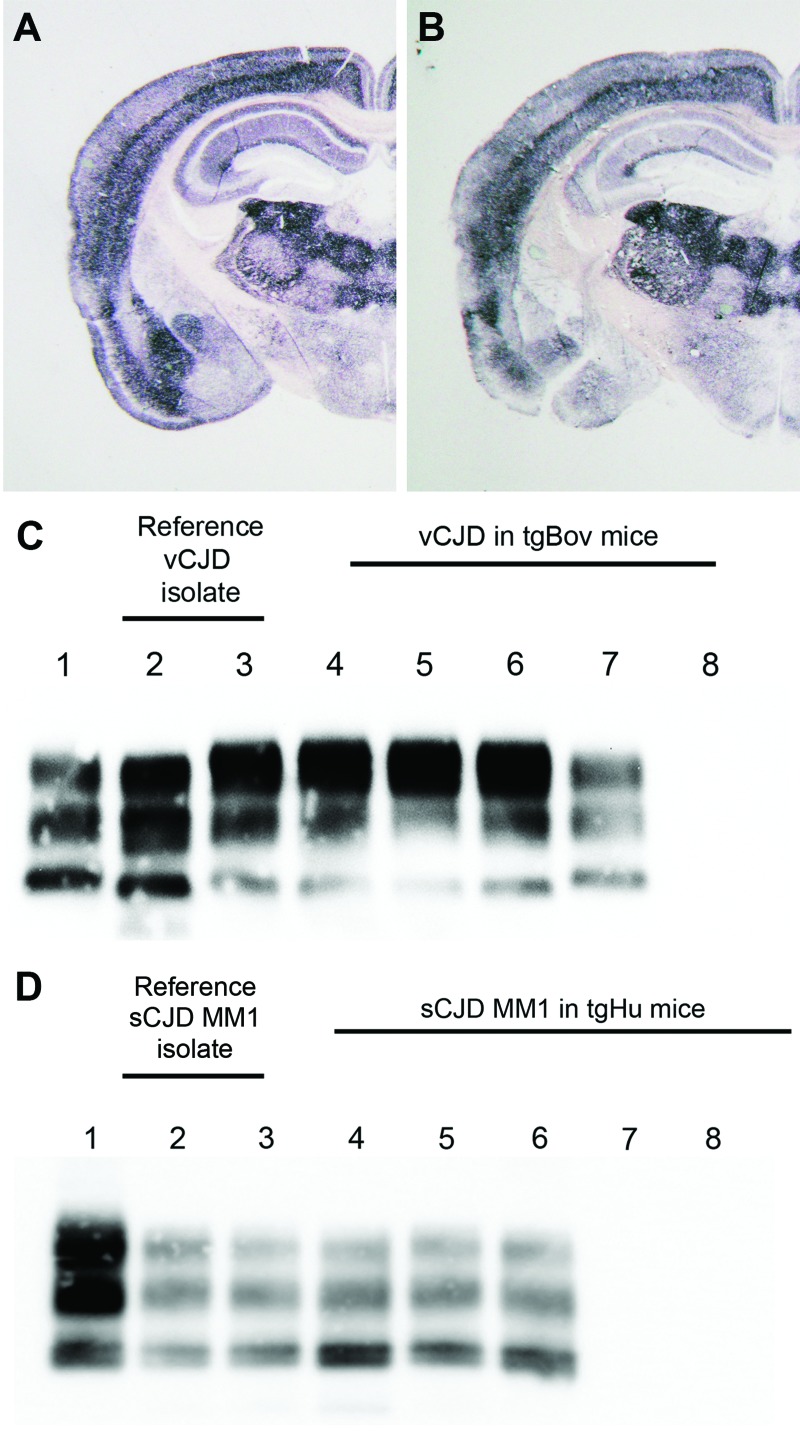
Figure.
Abnormal prion protein (PrPres) detection by using Western blot (WB) and paraffin-embedded tissue (PET) blot in the brain of transgenic mice expressing the methionine 129 variant of the human prion protein (PrP) (tgHu) or bovine PrP (tgBov). A, B) PET blot PrPres distribution in coronal section (thalamus level) of tgHu mice inoculated with sporadic Creutzfeldt-Jakob disease (sCJD) MM1 isolates (10% brain homogenate): A) reference isolate used for the endpoint titration in Table 1; B) sCJD case 1 (Table 2). C) PrPres WB of variant Creutzfeldt-Jakob disease (vCJD) reference isolate (used for endpoint titration in Table 1) and tgBov mice inoculated with the same vCJD reference isolate or vCJD blood fractions. Lane 1, WB-positive control; lanes 2 and 3, reference vCJD isolate; lane 4, leukocytes; lane 5, erythrocytes; lane 6, plasma; lane 7, WB-positive control; lane 8, healthy human plasma in tgBov. D) PrPres Western blot of the sCJD reference isolate (used for endpoint titration in Table 1) and tgHu mice inoculated with the same sCJD reference isolate and plasma from sCJD cases. A proteinase K–digested classical scrapie isolate in sheep was used as positive control for the blots in panels C and D. PrPres immunodetection in PET and Western blots was performed by using Sha31 monoclonal antibody (epitope: 145YEDRYYRE152 of the human PrP). Lane 1, WB-positive control; lanes 2 and 3, reference sCJD MM1 isolate; lane 4, brain tissue from case 1; lane 5, plasma from case 1; lane 6, plasma from case 3; lane 7, plasma from case 2; lane 8, plasma from case 4.

