
Figure 1.
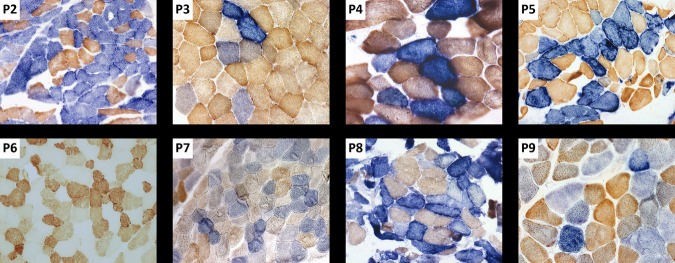
Sequential COX–SDH histochemistry. Sequential cytochrome c oxidase (COX) and succinate dehydrogenase (SDH) histochemistry was performed on skeletal muscle biopsies from all patients with the exception of patients 1 and 6. A mosaic pattern of COX activity is visible in each of the images, with COX-deficient fibers shown in blue and COX-positive fibers shown in brown. For patient 6, the individual COX histochemical reaction demonstrates a large number of COX-deficient fibers.