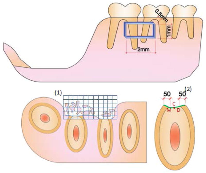
Figure 1. Schematic illustration of the assessed histomorphometric parameters.

Histomorphometry was used to assess percentage of defect filling (DF) (%), proportion of mineralized tissue (BD) (%), extension of new cementum (ENC) (μm), proportion of cementum-denuded root coverage by new cementum (PRC) (%) and thickness of new cementum (TNC) (μm). DF and BD were measured using a square grid (1) overlaid on the defect area. ENC was obtained by linear measurement of newly formed cementum, and PRC determined by the proportion of newly formed cementum covering the total extent of the instrumented root (green line). TNC measurements were made at fixed distances of 50 μm from mesial (M) and distal (D) side of pre-existing cementum, and on the central (C) region of the instrumented root (2).
