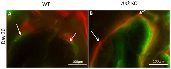
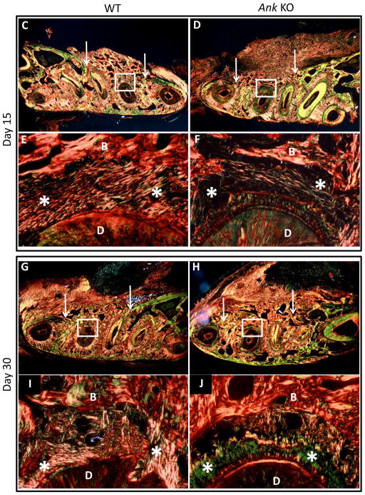
Figure 4. Labeling of new cementum apposition and periodontal ligament fiber organization.

(A–B) In regions of pre-existing cementum and bone away from the defect area, calcein dye (green) injected 7 days after surgery was observed with similar intensity in WT and Ank KO. Alizarin labeling (red) injected at day 14 was observed in WT and KO, though staining in Ank KO appear thicker and more concentrated than in controls. (C–J) Low magnification photographs of the surgically-created periodontal defects in the mandibles. Arrows indicate margins of defect and rectangles define the instrumented root area that is presented in higher magnification. Note that at 15 days post-surgery, both groups featured new bone formation (woven-like bone) with a disorganized connective tissue interfaced between the tooth and the new bone. At day 30, bone was more mature-like with regions of organized, parallel and functionally oriented PDL fibers identified in both WT and KO - Original magnification: figures C, D, G and H: 200x and figures E, F, I and J: 400x. Abbreviations: B: bone; D: dentin, star: periodontal ligament.

