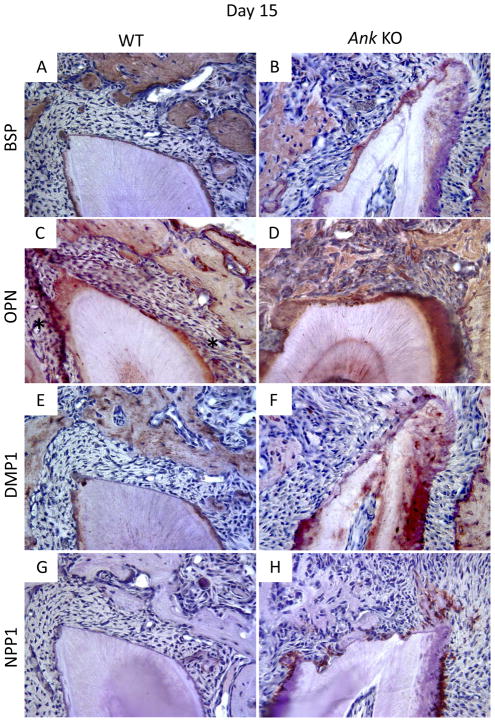
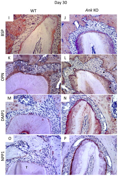
Figure 5. Expression of mineralized tissue markers during cementum regeneration.
Histological sections from days 15 and 30 were used for IHC. (A–B/I–J) Bone sialoprotein (BSP): By day 15, BSP protein expression was clear in the new bone of WT and KO mice, though little BSP was localized to the healing root surface. By day 30, the thin layer of new cementum on WT and KO roots featured positive BSP staining. (C–D/K–L) Osteopontin (OPN): OPN was positive in bone and cementum of WT and KO. By day 15, OPN was localized to the healing root surfaces in both WT and Ank KO molars, though more intense OPN staining was consistently identified in KO new cementum and PDL. OPN staining was similar in WT vs. KO at 30 days, except for notably increased OPN in Ank KO PDL. (E–F/M–N) Dentin matrix protein 1 (DMP1): DMP1 localized primarily to bone matrix around osteocytes, with lower levels of staining apparent in dentinal tubules. WT acellular cementum did not stain positively for DMP1. At day 15, there was no detectable DMP1 staining in healing sites either group. By day 30, DMP1 localized strongly to matrix and cells in Ank KO repair cementum, while in WT, DMP1 stained weakly and primarily around cementocytes. (G–H/O–P) Ectonucleotide pyrophosphatase phosphodiesterase 1 (NPP1): By day 15, no NPP1 expression could be detected at the healing site in WT samples, though in Ank KO, NPP1 expression was strong in cementoblasts associated pre-existing cementum. By day 30, NPP1 was detectable in some WT cementoblasts and osteoblasts on regenerated tissues. Osteoblast NPP1 expression in Ank KO was similar to WT, though cementoblasts at the healing site exhibited increased NPP1 -Original magnification: 400x.


