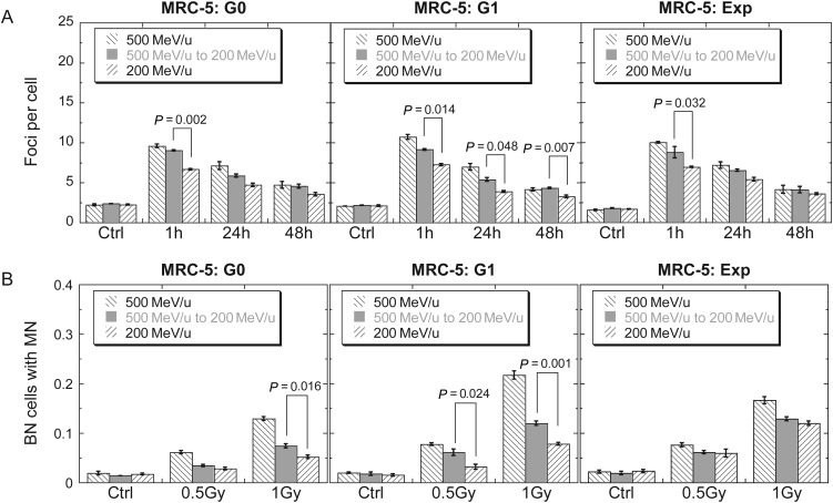
Fig. 3.
Secondary effects of PMMA on G0, G1 and exponentially growing MRC-5 cells. (A) Kinetics of 53BP1 foci in cells exposed to 0.5 Gy iron ions. (B) Binucleated cells (BN) with micronuclei (MN), which were obtained with a cytochalasin B-blocked micronucleus assay. Data were presented as mean ± SE. Experiments were independently repeated at least three times. P-values for comparisons between IR-A and IR-B were shown.

