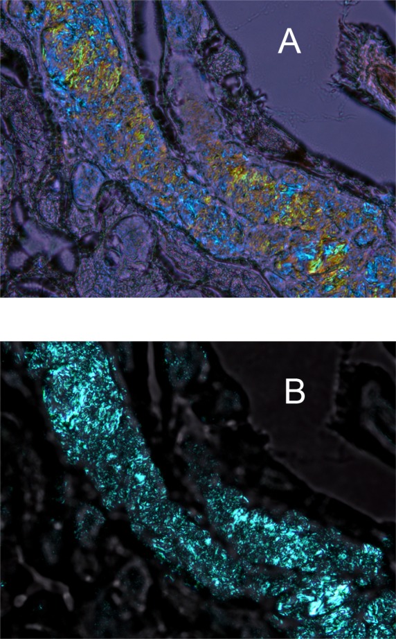
Figure 5.

Birefringence patterns of a cryosection of a mouse eyelid at room temperature. (A) Mouse meibomian gland as it is seen in cross-polarized light with the compensator in the light path. Note the microgranular structure of the lipid material within the meibomian duct. Different colors (from yellow to blue) indicate liquid-crystal zones with different predominant orientations of lipid molecules and/or different types of lipids. Magnification: ×400. (B) The same cryosection with no compensator. The contrast of the photograph is much higher than that in (A), but no conclusions can be made with regard to the presence or absence of zones with different predominant orientations (or directors) of lipid molecules, as no color information is available in this mode. Magnification: ×400.
