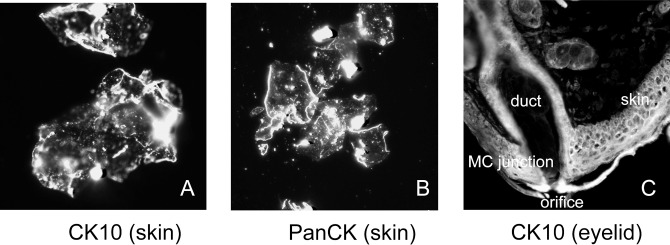
Figure 12.

Immunohistochemical anti-CK10 and anti-PanCK staining of particles of stratum corneum collected from human skin and anti-CK10 staining of a tissue section of a mouse eyelid. (A) Scrapings of human stratum corneum stained with anti-CK10 antibodies. (B) Particles of human stratum corneum stained with anti-PanCK antibodies. (C) An OCT-embedded 12-μm cryosection of a mouse eyelid stained with anti-CK10 antibodies. All pictures were taken with a magnification factor of 400 and cropped to approximately 250 × 250 μm.
