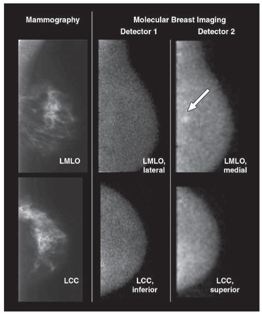
Fig. 3.
70-year-old woman with 6-mm invasive ductal carcinoma (arrow) in upper inner left breast initially identified on mammography as BI-RADS category 4 lesion. Screening mammograms and craniocaudal and mediolateral oblique views from both molecular breast imaging detectors are shown. When inferior and lateral molecular breast imaging views acquired with detector 1 (middle images) were reviewed in blinded reading session, they were interpreted as showing negative findings. When both inferior and lateral views from detector 1 and superior and medial molecular breast imaging views from detector 2 (right images) were available for interpretation, cancer was identified in medial view alone. Average uptake score of lesion visualized with molecular breast imaging was 3 (moderate focal uptake). LMLO = left mediolateral oblique, LCC = left craniocaudal.

