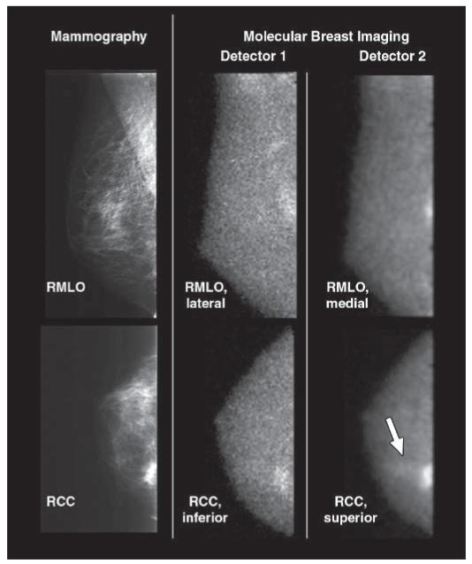
Fig. 4.
46-year-old woman with two foci of mixed invasive ductal carcinoma with ductal carcinoma in situ in upper inner right breast: One is located at 1-o’clock position 7 cm from nipple and second is located at 2-o’clock position 3 cm from nipple. Cancers were initially identified on mammography and sonography as BI-RADS category 5 lesions. Screening mammogram and craniocaudal and mediolateral oblique views from both molecular breast imaging detectors are shown. During blinded readings, when inferior and lateral molecular breast imaging views acquired with detector 1 (middle images) were available for interpretation (corresponding to views from single-head molecular breast imaging), one focus of cancer was detected. When superior and medial molecular breast imaging views from detector 2 were available in addition to detector 1 views during separate blinded reading session, same cancer was identified but average lesion uptake score was increased from 3 to 5, indicating increase in reader confidence in identifying lesion. Also, with additional detector 2 views, second focus of cancer (arrow) was identified as blush of low-intensity uptake (uptake score of 2) visible in superior craniocaudal view only. RMLO = right mediolateral oblique, RCC = right craniocaudal.

