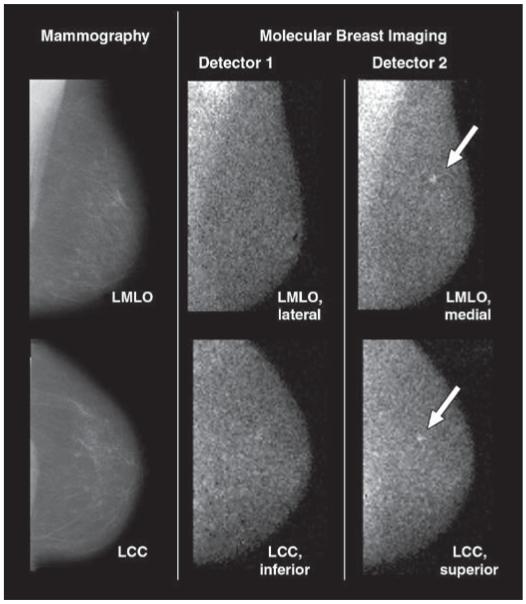
Fig. 5.
82-year-old woman with 4-mm invasive lobular carcinoma (arrows) in upper mid left breast at 12-o’clock position 6 cm from nipple. Cancer was initially identified on mammography as BI-RADS category 5 lesion; 5-mm benign intramammary lymph node in lower inner breast was also identified on mammography and sonography. Screening mammograms and craniocaudal and mediolateral oblique views from both molecular breast imaging detectors are shown. During blinded readings of only inferior and lateral molecular breast imaging views from detector 1 (middle images), molecular breast imaging findings were interpreted as negative. When superior and medial views from detector 2 (right images) were available for molecular breast imaging interpretation in addition to detector 1 views, cancer was identified and given uptake score of 4 (strong focal uptake) by two readers and score of 3 (moderate focal uptake) by third reader. Findings were otherwise negative on molecular breast imaging. LMLO = left mediolateral oblique, LCC = left craniocaudal.

