
Figure 7.
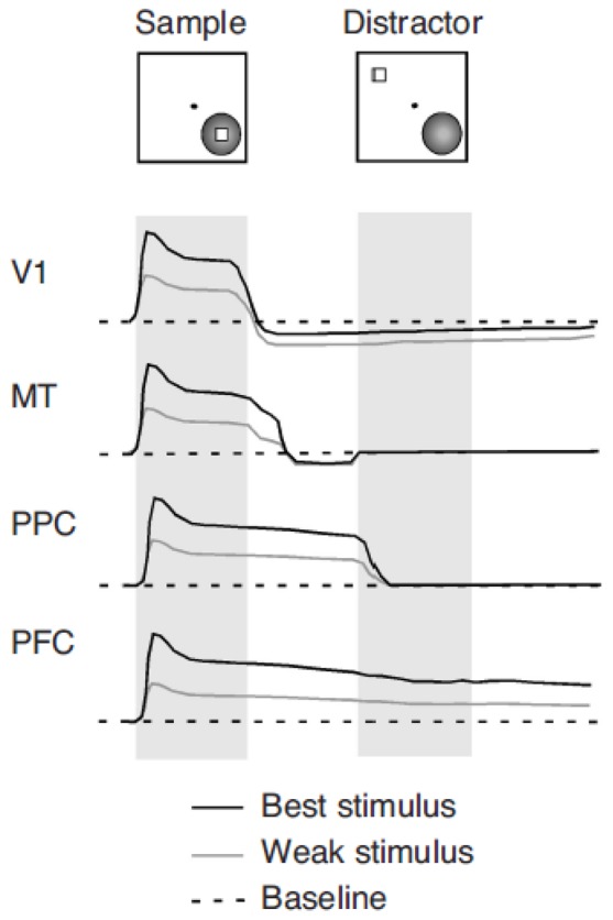
Manifestations of memory-related activity in successive visual cortical areas. Idealized neuronal responses are shown in response to two stimuli separated by a delay period: a sample that the subject is required to remember and a subsequent distractor. The black line represents responses to an optimal stimulus evoking the best response from the neuron. The gray line represents responses to a less effective stimulus evoking a moderate neuronal response. The dotted line represents the neurons’ discharge rate baseline. Neuronal discharges in a small percentage of V1 neurons recede below the baseline during the active maintenance in memory of an optimal stimulus. Neurons in MT exhibit a short-lived persistent discharge that follows the disappearance of the stimulus. Their activity quickly returns to baseline, however. Neurons in posterior parietal cortex (PPC)—for example, in areas 7a and LIP—respond with sustained discharges in the delay period following the sample. However, these are terminated by the appearance of a distracting stimulus, outside the receptive field. Neurons in prefrontal cortex (PFC)—for example, in areas 8a and 46—respond with sustained responses to the stimulus, which are not interrupted by the appearance of the distractor.