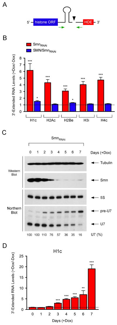
Figure 2. SMN is required for 3′-end formation of histone mRNAs.
(A) Schematic of the 3′-end structure of histone mRNAs and position of RT-qPCR primers. (B) RT-qPCR analysis of 3′-extended histone mRNAs in NIH3T3-SmnRNAi and NIH3T3-SMN/SmnRNAi cells cultured with or without Dox for 5 days. RNA levels in Dox-treated cells were expressed relative to untreated cells (dashed line). (C) Temporal analysis of SMN and U7 levels in NIH3T3-SmnRNAi cells cultured with Dox for the indicated number of days. (D) RT-qPCR analysis of the time-dependent accumulation of 3′-extended histone mRNAs in NIH3T3-SmnRNAi cells cultured as in (C). RNA levels in Dox-treated cells were expressed relative to untreated cells (dashed line).
Data in all graphs are represented as mean and SEM.

