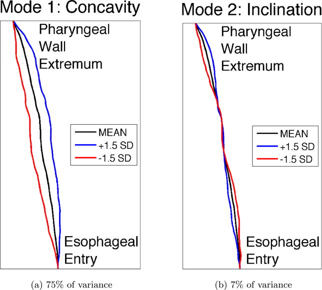
Figure 6.

The two largest modes of variation in posterior pharyngeal wall shape, determined in completely data-driven fashion, without imposing any prior notions about expected shape variations, by applying PCA to the observed pharyngeal wall shapes from the subject pool. Modes reflect differences in concavity and inclination of the pharyngeal wall. The overall mean pharyngeal wall shape is shown in black, and the blue and red lines show the nature of deviations from the mean according to each mode. The magnitude of the deviations shown reflect the magnitude of variations seen in the subject pool, at precisely +/- 1.5 standard deviations from the mean shape. Because these modes account for over 82% of the overall variance, arbitrary pharyngeal wall shapes may be well represented using only these two modes.
