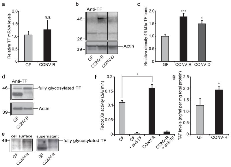
Figure 2. The gut microbiota increases TF procoagulant activity and cell-surface localization.
a, Relative levels of mRNA for TF in sections of small intestine from GF and CONV-R mice (n = 7–11 mice per group). b, Anti-TF immunoblot of small-intestinal lysates from GF, CONV-R and CONV-D mice. c, Quantification of the 46-kDa TF band shown in b (n = 14–25 mice per group). Data are normalized to actin and expressed relative to GF. d, Anti-TF immunoblot of primary enterocytes (from GF and CONV-R mice) after 2 h of culture. e, Anti-TF immunoblots from N-hydroxysuccinimido-biotin-labelled primary enterocytes from GF and CONV-R mice. Left: pull-down of proteins located on the plasma membrane with NeutrAvidin beads. Right: supernatant containing unlabelled proteins. f, Factor Xa activity in small-intestinal lysates from GF and CONV-R mice treated with control or anti-TF antibody (n = 4 or 5 mice per group). g, Levels of thrombin–antithrombin (TAT) complexes in small-intestinal lysates from GF and CONV-R mice (n = 7 mice per group). Female Swiss Webster mice or cells isolated from these mice were analysed in all panels. Results are shown as means ± s.e.m. Asterisk, P < 0.05; three asterisks, P < 0.005; n.s., not significant.

