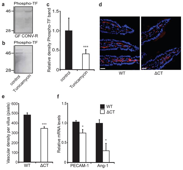
Figure 3. The gut microbiota increases phosphorylation of the cytoplasmic tail of TF, which increases vessel density in the intestine.
a, b, Anti-phospho-TF immunoblot of (a) small-intestinal lysates from GF and CONV-R mice and (b) primary enterocytes (from CONV-R mice) incubated for 2 h in the absence and presence of tunicamycin (10 μmol l−1). c, Quantification of b (n = 5 mice per group). d, PECAM-1 staining (red) of sections of small intestine from 10–12-week-old wild-type (WT) and ΔCT female mice on a C57Bl6/J genetic background. Nuclei were stained with Hoechst nuclear dye (blue). e, Quantification of d (n = 4–6 mice per group). f, Relative levels of mRNA for PECAM-1 and Ang-1 in segments of small intestine from WT and ΔCT mice (n = 3 or 4 mice per group). Female Swiss Webster mice or cells isolated from these mice were analysed in a–c. Scale bars, 20 μm. Results are shown as means ± s.e.m. Asterisk, P < 0.05; three asterisks, P < 0.005.

