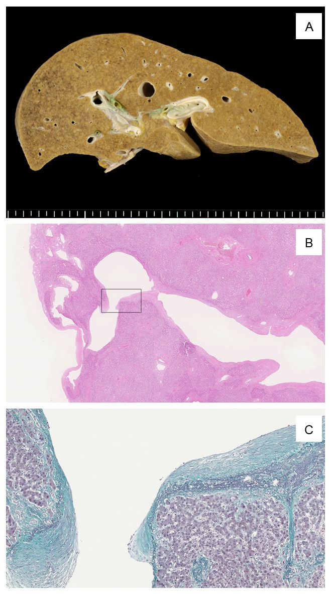
Figure 2.

A: Liver shows a little irregular congestion and slight granular appearance macroscopically. B: Some distended shunt vessels were identified histologically in both lobes of the liver. This image shows one of those vessels. This vessel was located directly under the serosa aside from venous ligament. Box indicates the site of image C. C: Shunt vessel was accompanied by incrassate elastic lamellae (Elastica-Masson stain).
