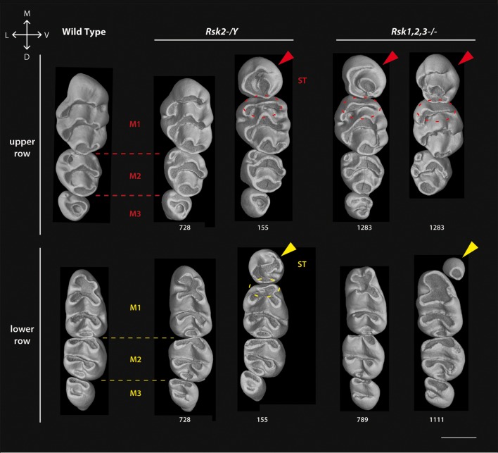
Figure 3. Variation of molar shape, size and number in Rsk2-/Y and Rsk1,2,3-/- mice analyzed by X-Ray microtomography.
All molar rows are oriented in the same manner (top corresponds to mesial and left to lingual side). At the left are wild-type molars, other rows are mutants as indicated. Arrowheads point to the supernumerary teeth ST; dotted ellipses show the reduction of the mesial-most affected cusp. Scale bar: 0.7 µm.

