
Fig.7. The conserved ULK phosphorylation site in C. elegans Bec-1 is required for autophagy.
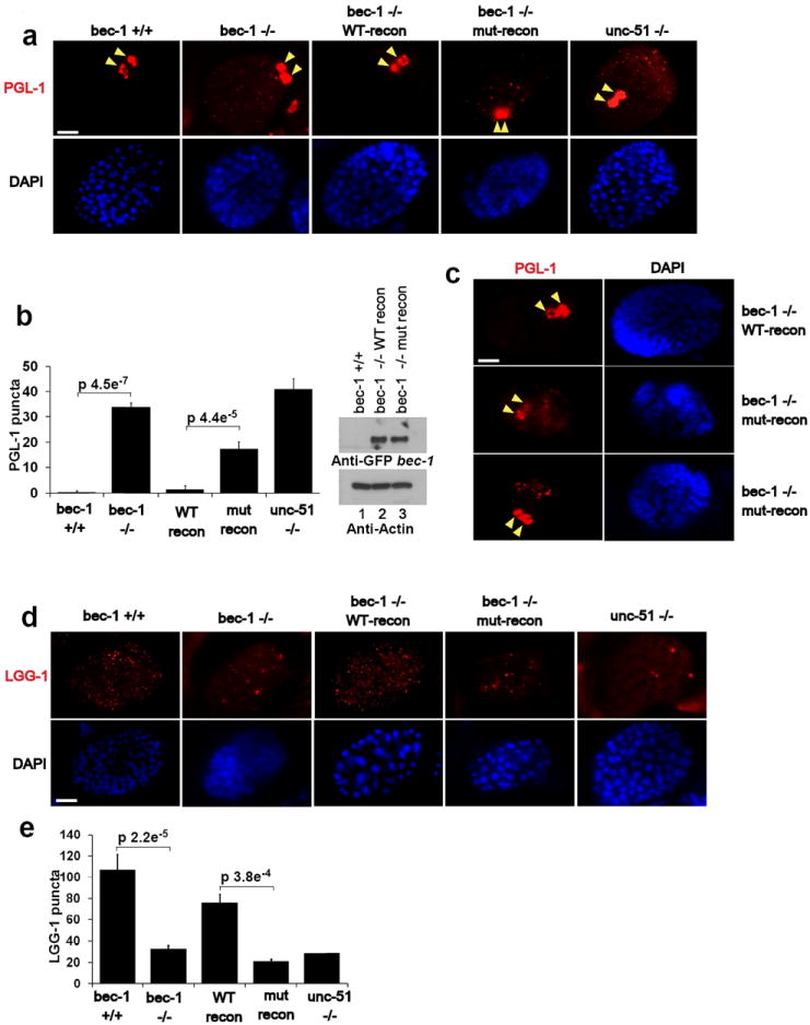
Unless otherwise stated all experiments were repeated three times and data shown is representative. (a) Bec-1 C. elegans were reconstituted with either wild-type or mutant GFP-BEC-1. Stable worm lines with Bec-1 rescue were obtained and embryos were stained with anti-PGL-1 antibody. Arrow indicates normal PGL-1 staining in germline cells. Scale bars represent 10μm. (b) Quantification of PGL-1 puncta outside germline cells (left panel). Error bars represents standard deviation between 3 unique embryos in a representative experiment. Reconstituted Bec-1 (WT and mut) levels in Bec -/- stable worms were compared by Western blot. Mean value presented. (c) Spectrum of defects in PGL granule degradation in bec-1 mutant rescue embryos. Mutant embryos displayed either high levels of diffuse PGL-1 staining (middle-left panel, 1/3 of the embryos), or large punctuate PGL-1 structures in somatic cells (bottom-left panel. 2/3 of the embryos). Both diffuse or punctuate PGL-1 staining in somatic cells have been described in autophagy deficient embryos. (d) Embryos from the lines described in Fig.6a were labeled with anti-LGG-1, along with wild-type and unc-51 worms. Representative embryos at ~100 cell stage are shown. (e) Quantification of LGG-1 per embryo from labeling in panel d. Error bars generated as in panel b. Mean value presented.