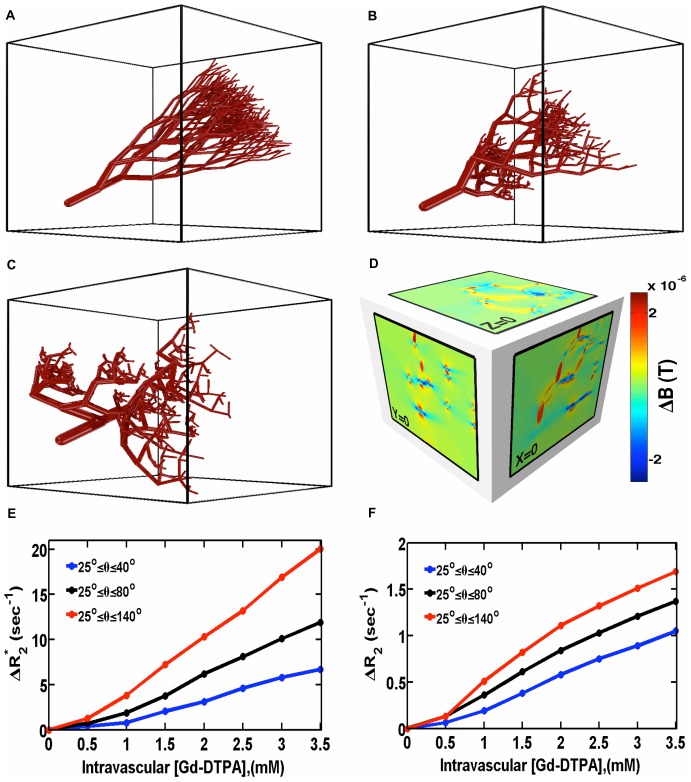
Figure 4. The influence of vascular morphology on ΔR2 * and ΔR2.
(a–c) Sample microvascular networks simulated using a fractal tree model with increasing branching angle heterogeneity. (d) Three orthogonal slices through the magnetic field perturbation at the body center for the vascular network in (c). (e–f) Effect of branching angle heterogeneity on the concentration dependence of ΔR2 * and ΔR2 computed with FPFDM (B0 = 4.7T, Δχ = 1×10⁻⁷, 2% target vascular volume fraction). Both ΔR2 and ΔR2 * increase with branching angle heterogeneity.

