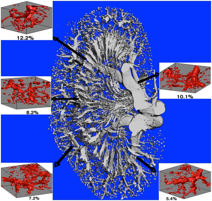
Figure 7. Kidney vascular structure extracted from micro-CT.
Kidney vasculature extracted from micro-CT along with representative MR voxel-sized (1 mm3) microvascular models taken from different sections of the kidney vasculature with their respective vascular volume fractions. The existence of the bubble-like structures demonstrates the filling of glomeruli with Microfil but a higher resolution would be required to differentiate the individual capillaries.

