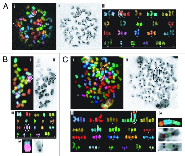
Figure 4. Mice with conditional inactivation of Tp53 initiating in HSCs or DN thymocytes develop lymphomas with oncogenic antigen receptor locus translocations. (A) Cytogenetic analysis of a metaphase from VP lymphoma no. 820 with the clonal t(2;17) translocation circled or isolated. (A, i) Spectral image. (A, ii) DAPI image. (A, iii) Karyotype table. (B) Cytogenetic analyses of a metaphase or chromosome from VP lymphoma no. 421 with the clonal t(12;15) translocation circled. (B, i) SKY image. (B, ii) DAPI image. (B, iii) Karyotype table. (B, iv). SKY (left) or FISH image (right) of the t(12;15) translocation hybridized with 3′IgH (green) and c-Myc (red) probes. (C) Cytogenetic analyses of a metaphase or chromosome from LP lymphoma no. 902 with the clonal t(4;15;14) translocation circled or isolated. (B, i) SKY image. (B, ii) DAPI image. (B, iii) Karyotype table. (B, iv) SKY (top) or FISH images of the t(4;15;14) translocation. (middle) hybridized with TCRVδ3/Vα6 (green) and TCRCα (red) probes. (bottom) hybridized with c-Myc (green) probe.

An official website of the United States government
Here's how you know
Official websites use .gov
A
.gov website belongs to an official
government organization in the United States.
Secure .gov websites use HTTPS
A lock (
) or https:// means you've safely
connected to the .gov website. Share sensitive
information only on official, secure websites.
