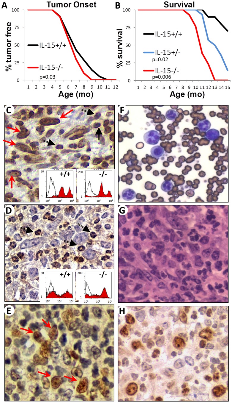
Figure 1. IL-15 is not required for Tax Tumorigenesis.
A) Incidence of tumor onset in IL-15−/− TAX-LUC mice (red line, n = 10) compared to IL-15+/+ TAX-LUC littermate control mice (black line, n = 21), p = 0.03 (2-tailed, paired Student’s T Test). B) Survival curve comparing IL-15−/− TAX-LUC mice (red line, n = 13) and IL-15+/− TAX-LUC mice (blue line, n = 14) to IL-15+/+ TAX-LUC littermate control mice (black line, n = 10). p = 0.006 for KO vs. WT and p = 0.02 for HET vs. WT. C) Image of CD16/32 immunohistochemistry of IL-15−/− TAX-LUC tumor sections in which the malignant large granular lymphocytes (red arrows) are stained along with an admixture of tumor infiltrating neutrophils (black arrows). Insets are FACS histograms of homogenates from IL-15+/+ TAX-LUC or IL-15−/− TAX-LUC tail tumors unstained (white curves) or stained with anti-CD16/32 FITC (red curves). D) Image of Ly6G immunohistochemistry of IL-15−/− TAX-LUC tumor sections in which tumor infiltrating neutrophils (black arrows) are stained. Insets are FACS histograms of homogenates from IL-15+/+ TAX-LUC or IL-15−/− TAX-LUC tail tumors unstained (white curves) or stained with anti-Ly6G-PE (red curves). E) Image of phosho-RelA immunohistochemistry of IL-15−/− TAX-LUC tumor sections in which many of the malignant large granular lymphocytes (red arrows) show nuclear localization as a marker of NFκB activation. F) Image of a peripheral blood smear (Wright’s stain). G) Hematoxylin and eosin stained IL-15−/− TAX-LUC tumor section. H) Image of Ki67 immunohistochemistry of IL-15−/− TAX-LUC tumor sections.

