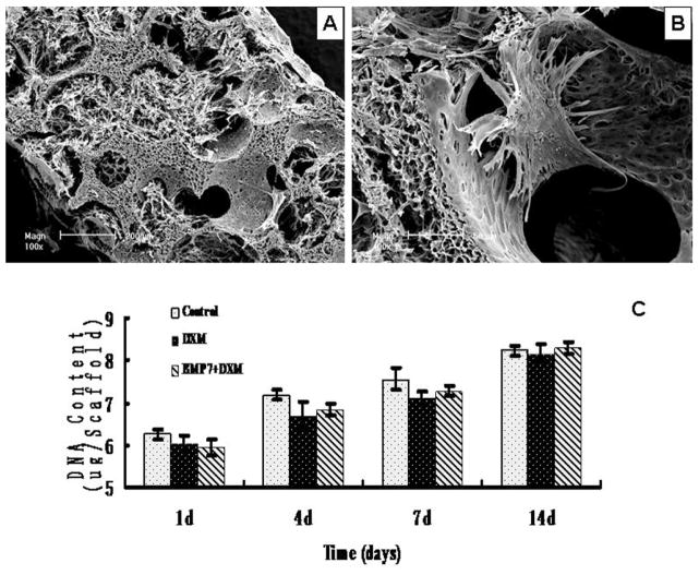
Fig. 2.
(A) SEM observation of NF-PLLA scaffolds loaded with DPSCs cultured in vitro for 3 days at a low magnification. The interconnected macro pores in the scaffold were filled with DPSCs, which had long cell processes and secreted abundant ECM. (B) SEM observation of NF-PLLA scaffolds loaded with DPSCs cultured in vitro for 3 days at a higher magnification. The DPSCs attached and fully spread on the pore surfaces of the NF-PLLA scaffolds. (C) The proliferation characteristics of DPSCs cultured on NF-PLLA scaffolds in different media.

