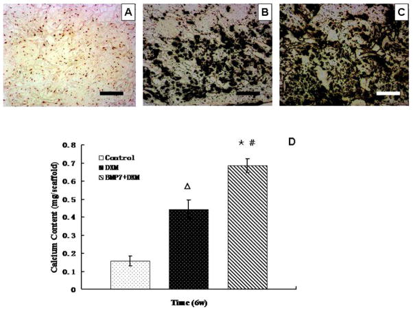
Fig. 6.
von Kossa staining views of DPSCs on NF-PLLA scaffolds cultured for 6 weeks in vitro (dark color indicates mineral nodules): (A) control group; (B) “DXM” group; (C) “BMP-7 + DXM” group. (D) Calcium content quantification of DPSCs on NF-PLLA scaffolds cultured for 6 weeks in vitro. The bars represent means ± SD (n = 3). “BMP-7 + DXM” group compared to control group: *p<0.05; “BMP-7 + DXM” group compared to “DXM” group: #p<0.05; “DXM” group compared to control group: Δp<0.05. Scale bars represent 100 μm. Original magnification: ×20.

