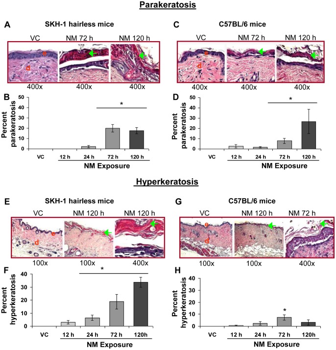
Figure 3. NM exposure causes parakeratosis and hyperkeratosis in the skin epidermis of SKH-1 and C57BL/6 mice.
Dorsal skin of mice was exposed topically to either 200 µL of acetone or NM (3.2 mg) in 200 µL acetone. After 12, 24, 72 and 120 h of NM exposure, mice were sacrificed and dorsal skin tissue sections (5 µM) were processed, H&E stained and analyzed as detailed under Materials and Methods. Panels A and C (i–iii) and E and G (i–iii) are representative H&E stained skin sections (100 or 400× magnification) showing parakeratosis and hyperkeratosis, respectively, from vehicle control as well as NM exposed (72 and 120 h) skin tissue in mice. These NM-related histopathological changes were assessed as detailed under materials and methods, and calculated as percent length of mice skin epidermis showing parakeratosis (B and D) and hyperkeratosis (F and H). Data presented are mean ± SEM of 3–5 animals in each group. *, p<0.05 compared to respective vehicle control; VC, vehicle control; NM, nitrogen mustard; e, epidermis; d, dermis; green arrows, parakeratosis, hypercornification or acanthosis.

