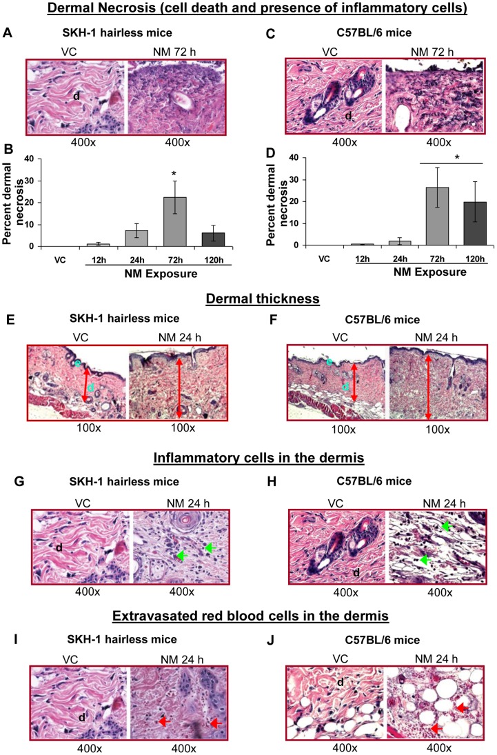
Figure 5. NM exposure causes necrosis, dermal thickness, recruitment of inflammatory cells and extravasated red blood cells in the dermal region of SKH-1 and C57BL/6 mice skin.
Dorsal skin of mice was exposed topically to either 200 µL of acetone or NM (3.2 mg) in 200 µL acetone. After 12, 24, 72 and 120 h of NM exposure, mice were sacrificed and dorsal skin tissue sections (5 µM) were processed, H&E stained and analyzed as detailed under Materials and Methods. Panels A and C, E and F, G and H, and I and J are representative H&E stained skin sections from vehicle control as well as NM exposed skin tissue in mice for dermal necrosis (with presence of breaking down inflammatory cells, mainly neutrophils), dermal thickness, inflammatory cells, and extravasated red blood cells. These NM-related histopathological changes were assessed as detailed under materials and methods, and calculated as percent area of mice skin dermis showing necrosis (B and D), inflammatory cells (Table 1), and extravasated red blood cells (Table 2). *, p<0.05 compared to respective vehicle control; VC, vehicle control; NM, nitrogen mustard; d, dermis; green arrows, inflammatory cells; red arrows, extravasated red blood cells.

